| Anatomical Chart Company |
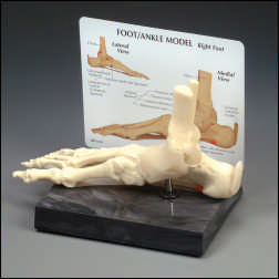 |
| Foot and Ankle Model |
|
Life-size model of a right foot features the platar calcaneonavicular (spring) ligament with plantar fascitis. Also includes tibia, fibula, calcaneus,... |
|
| Anatomical Chart Company |
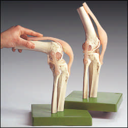 |
| Functional Model of the Knee Joint |
|
This plastic model of the right knee features an exclusive detachable ligament system allowing a complete view of each bone, ligament and cartilage. T... |
|
| Anatomical Chart Company |
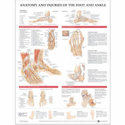 |
| Anatomy and Injuries of the Foot and Ankle Chart |
|
Shows medial and lateral view of the bones and ligaments of the foot and ankle. Illustrates nerve and blood supply to this region, including plantar v... |
|
| Anatomical Chart Company |
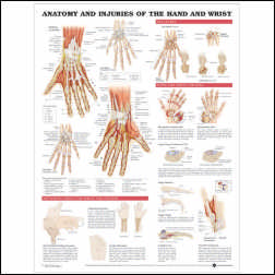 |
| Anatomy and Injuries of the Hand and Wrist Anatomical Chart |
|
Shows normal anatomy, including bones, ligaments, muscles, nerves and blood vessels. Illustrates and describes common acute fractures, such as Colles'... |
|
| Anatomical Chart Company |
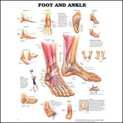 |
| Foot and Ankle Anatomical Chart |
|
Illustrates foot and ankle anatomy including bones, muscles and tendons. Shows medial, frontal, lateral, and plantar views as well as a cross section.... |
|
| Anatomical Chart Company |
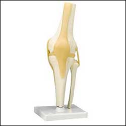 |
| Functional Knee Joint Model |
|
Life-size functional model demonstrates flexion, extension, and internal/external rotation. Includes flexible artificial ligaments. Life-size, removab... |
|
| Anatomical Chart Company |
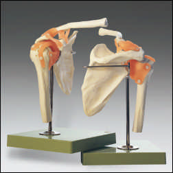 |
| Functional Model of the Shoulder Joint |
|
This life-size right shoulder joint features an exclusive detachable ligament system which allows individual viewing of each bone, ligament and cartil... |
|
| Anatomical Chart Company |
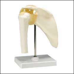 |
| Functional Shoulder Joint Model |
|
An instructional model to illustrate abduction, adduction, anteversion, retroversion, internal/external rotation. Includes flexible, artificial ligame... |
|
| Anatomical Chart Company |
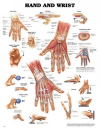 |
| Hand & Wrist Chart |
|
Rigid plastic lamination with metal eyelets in top corners for convenient wall hanging or for use with portable chart stands; mark able (write-on/wipe... |
|
| Anatomical Chart Company |
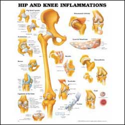 |
| HIp and Knee Inflammations Anatomical Chart |
|
Shows general hip and knee anatomy, as well as hip joint capsule, acetabulum, brusae, ligaments of the knee, and detailed anatomy of a tendon. Illustr... |
|
| Anatomical Chart Company |
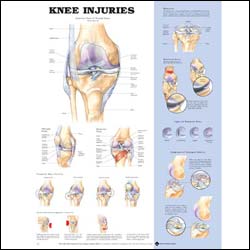 |
| Knee Injuries Chart |
|
Show anterior view of normal knee anatomy (patella removed), as well as oblique and posterior views. Illustrates traumatic knee injuries, meniscus, ty... |
|
| Anatomical Chart Company |
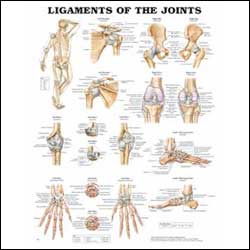 |
| Ligaments of the Joints Anatomical Chart |
|
Shows location of various joints provides anterior and posterior views of the left shoulder, right hip, right knee and left elbow. Also illustrates la... |
|
| Anatomical Chart Company |
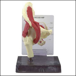 |
| Muscled Hip Joint Model |
|
Articulating right hip joint featrues the following muscles: gluteus, mediu, piriformis, gemellus superior and inferior with obturator internus and il... |
|
| Anatomical Chart Company |
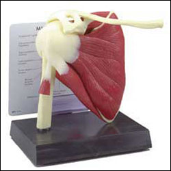 |
| Muscled Shoulder Joint Model |
|
Semi-articulating right rotator cuff model has an encapsulated humerus that is removable from the capsular ligament of the shoulder joint. This allows... |
|
| Anatomical Chart Company |
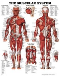 |
| Muscular System Chart |
|
Rigid plastic lamination with metal eyelets in top corners for convenient wall hanging or for use with portable chart stands; mark able (write-on/wipe... |
|
| Anatomical Chart Company |
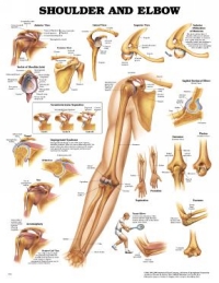 |
| Shoulder & Elbow Chart |
|
Rigid plastic lamination with metal eyelets in top corners for convenient wall hanging or for use with portable chart stands; mark able (write-on/wipe... |
|
| Anatomical Chart Company |
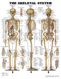 |
| Skeletal System Chart |
|
Rigid plastic lamination with metal eyelets in top corners for convenient wall hanging or for use with portable chart stands; mark able (write-on/wipe... |
|
| Anatomical Chart Company |
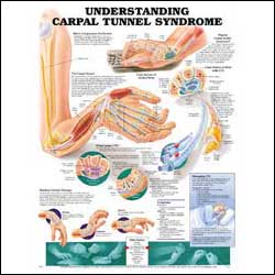 |
| Understanding Carpal Tunnel Syndrome Anatomical Chart |
|
Defines Carpal Tunnel Syndrome (CTS) and nerve compression syndrome. Shows the Carpal Tunnel and cross sections of a normal wrist and one with CTS. Ca... |
|
| Anatomical Chart Company |
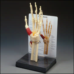 |
| Wrist and Hand Model |
|
Life size model of a left hand, wrist and forearm features: distal, middle and proximal phalanges of the thumb, metacarpal bones, joint capsule ligame... |
|
|

